Use this guide one step at a time.
Key points
- Review the pathology report for melanoma site, thickness, ulceration, margin status, and stage; ask whether expert review is needed for an unusual subtype.
- Ask whether sentinel lymph-node biopsy would add useful staging information and how a positive result would change surveillance or additional treatment.
- For higher-risk or advanced melanoma, ask whether BRAF testing is needed and compare immunotherapy, targeted therapy, local treatment, and trial options.
Subtypes and biomarkers
Cutaneous melanoma begins in skin melanocytes. Acral melanoma arises on palms, soles, or under nails; mucosal and uveal melanomas begin in different tissues and follow different testing and treatment pathways. Desmoplastic and other uncommon forms may also warrant specialist pathology review. For cutaneous melanoma, the pathology report should document tumor thickness, ulceration, margins, and other features used with lymph-node and distant-spread findings to determine stage. Sentinel lymph-node status can add prognostic and staging information for selected tumors. Molecular testing becomes especially relevant in higher-risk, unresectable, or metastatic disease. BRAF V600 alterations can make combined BRAF and MEK targeted therapy an option; other alterations may support a clinical-trial discussion. Immunotherapy does not require a BRAF alteration, and PD-L1 results do not function as a simple yes-or-no gate for standard melanoma immunotherapy. A skin primary should also be distinguished from mucosal or eye melanoma because those diagnoses are not interchangeable.
What is melanoma?
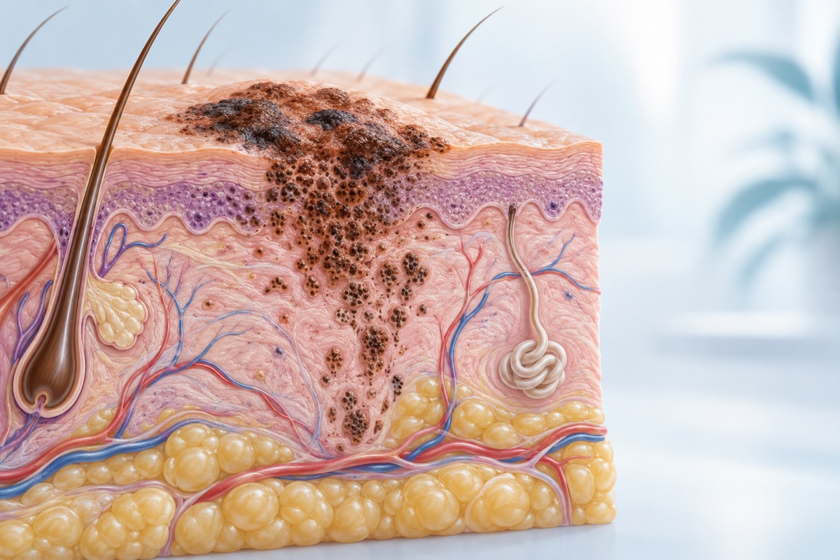
Most melanomas begin in skin, although melanoma can arise in the eye, mucosal surfaces, or other sites and is managed differently. Cutaneous melanoma develops when melanocytes acquire changes that permit uncontrolled growth. Ultraviolet exposure, including sun and tanning-device exposure, contributes to risk; fair skin, many or atypical moles, personal or family history, and certain inherited changes also increase risk, though anyone can develop melanoma. Clinicians classify melanoma by its microscopic features and whether it remains local, reaches regional lymph nodes, or spreads to distant organs. Tumor thickness and ulceration are especially important in localized disease. Molecular testing can identify changes, including BRAF alterations, that may guide therapy in advanced or selected high-risk disease. Because melanoma can recur, follow-up plans commonly include skin and lymph-node examinations, education about new or changing lesions, and imaging or laboratory testing when clinically indicated. A personal history also increases the chance of another primary melanoma, making continued whole-skin surveillance important.
Signs and symptoms
A melanoma may appear as a new spot or as a change in an existing mole or pigmented area. Warning features include asymmetry, an irregular border, varied colors, increasing diameter, and evolution over time. A lesion may itch, bleed, ooze, ulcerate, or look noticeably different from nearby moles. Some melanomas are pink, red, skin-colored, or have little pigment, so color alone cannot rule them out. Melanoma can also develop under a nail, on palms or soles, or in less visible areas. Later symptoms depend on where disease has spread and may include enlarged lymph nodes or organ-specific problems. These changes can also result from noncancer conditions. A clinician should evaluate a persistent, changing, bleeding, or otherwise concerning skin lesion rather than relying on photographs or self-assessment alone.
Diagnosis and staging
Evaluation usually begins with a complete skin examination and review of personal and family history. A suspicious lesion is removed or sampled for microscopic examination; an excisional biopsy is often preferred when feasible because the report must measure features such as melanoma thickness and ulceration. After melanoma is confirmed, the team may examine regional lymph nodes and consider sentinel lymph-node biopsy for tumors with sufficient risk. CT, PET, MRI, or other imaging is used selectively, particularly for symptoms or higher-stage disease. Staging describes the primary tumor, regional nodes, and distant metastases, grouped from stage 0 through IV. Pathology may also report mitotic activity and other features. Biomarker testing, including BRAF testing, is relevant for some higher-risk, unresectable, or metastatic melanomas. A pathology review may clarify an uncertain diagnosis or uncommon subtype before major treatment. Care depends on the melanoma site and subtype, stage, pathology, biomarkers, prior treatment, and the person’s health and goals.
Established treatment paths
Surgical excision with an appropriate margin is the main treatment for localized cutaneous melanoma. Sentinel lymph-node biopsy may be offered for staging when tumor features indicate a meaningful risk of nodal spread; routine extensive node removal is not required for every positive sentinel node. Some people with resected, higher-risk melanoma receive adjuvant immunotherapy, and selected BRAF V600–mutated melanomas may be treated with combined BRAF and MEK targeted therapy. For unresectable or metastatic disease, immune checkpoint inhibitors are central options, used alone or in combinations selected for disease burden, expected benefit, and toxicity. BRAF/MEK combinations are options when an actionable BRAF mutation is present and can produce rapid responses. Surgery or focused radiation may help selected sites, while radiation can also relieve symptoms. Treatment of melanoma in the eye or mucosal sites follows different pathways. Clinical trials may test new drug combinations, cellular therapies, or treatment sequences. Treatment response and immune-related toxicity require planned clinical, laboratory, and imaging follow-up. The plan depends on type, stage, biomarkers, prior therapy, pace and sites of disease, organ function, and patient preferences.
Supportive care
Supportive needs depend strongly on treatment. Lymph-node procedures can cause swelling; early lymphedema assessment may help. Immunotherapy can trigger inflammation in the bowel, lungs, liver, hormone glands, skin, nerves, or other organs. New diarrhea, cough, breathlessness, severe fatigue, headache, weakness, rash, or vision change warrants prompt contact rather than waiting for the next visit. Targeted therapy can cause fever, rash, fatigue, and other symptoms that need a treatment-specific plan. Dermatology follow-up, sun protection, and self-awareness help detect new lesions but do not treat melanoma. Counseling, social work, pain care, rehabilitation, and palliative care can accompany anticancer therapy.
Questions to ask your cancer care team
Use the questions that fit your situation, and add the details that matter most to you.
- What melanoma subtype, thickness, ulceration status, and stage are documented in my pathology report?
- Do I need sentinel lymph-node biopsy or additional imaging, and what would the results change?
- Should my tumor be tested for BRAF or other biomarkers before choosing treatment?
- What benefits, side effects, and monitoring needs come with each recommended option?
- What follow-up schedule and skin or lymph-node changes should prompt an earlier visit?
Clinics for people exploring melanoma care

Sunridge Medical Center
Sunridge Medical reports treating all advanced cancers. Its published materials specifically name melanoma care.
Featured placement is separate from an independent ranking or evidence that a clinic is the best fit for a particular patient.Other source-matched clinic profiles
These profiles mention a matching cancer specialty in their published materials. Browse their locations, reported services, and practical details.



Sources
Source information checked: 2026-10-01. The links below identify the public and clinic-provided materials used for this page.
- NCI: Melanoma Treatment (Patient Version) ↗Checked 2026-09-29
- NCI: Skin Cancer, Including Melanoma ↗Checked 2026-09-29
- NCI: Side Effects of Cancer Treatment ↗Checked 2026-09-29
- NCI: Sentinel Lymph Node Biopsy ↗Checked 2026-10-01
- NCI: Immunotherapy Side Effects ↗Checked 2026-10-01
- NCI: Lifileucel ↗Checked 2026-10-01
How Cancer Clinic Finder compiles information · Suggest a factual correction